Floor Of Left Maxillary Sinus
Panoramic Radiograph Of The Case Opacification Of Left Maxillary Download Scientific Diagram
Benign Maxillary Sinus Masses Ento Key

Cbct Images Of Left Maxillary Sinus In Sagittal View Depicting Relation Download Scientific Diagram

Thick Inspissated Mucous Covering The Left Maxillary Sinus Floor Download Scientific Diagram
Radiology Of Maxillary Sinus

Isolated Unilateral Upper Alveolar Numbness In Silent Sinus Syndrome Bmj Case Reports

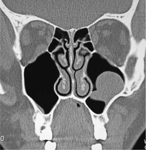
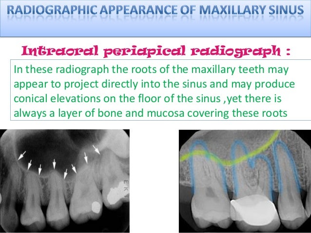
Citation needed projecting into the floor of the antrum are several conical processes corresponding to the roots of the first and second maxillary molar teeth.
Floor of left maxillary sinus. My brother s cemri shows diffuse mucosal thickening are seen in both ethmoidal both maxillary sinuses more on left side answered by dr. Inside has a mucous film that almost does not feel the elements due to the lack of nerve and blood vessels. When a ct scan is taken of the head the sinuses should show up black since they are cavities. Of the symptoms can be identified random sudden single discharge from one side of the nose.
Causes the sinuses to be swollen. Because the symptoms manifest themselves in a serious complication of the disease. When a tooth is lost the alveolar process. When the area shows up white or gray it is called opaque or opacification of the sinus.
The maxillary sinus is the cavity behind your cheeks very close to your nose 1. Within the maxillary sinus which lies beneath the cheek bone on each side are mucous glands a blockage in the mucous duct can cause the gland to enlarge which can lead to the formation of a dome shaped maxillary mucous retention cyst the cyst does not usually cause any symptoms and does not damage expand or thin the wall of the sinus. The ostia of the maxillary sinus often clog because the ostia are. If the sinus is large it reaches below this level.
As there is no normal tissues regeneration and the excretory ducts patency of the mucous glands is not restored. Cyst of left and right maxillary sinus. A maxillary sinus cyst is an abnormal tissue growth located in either of the cavities located behind the cheekbones on either side of the nose. These cavities are called sinuses and they are located in the maxilla or upper jaw cysts are closed pocket like formations of tissue and are filled with liquid air or semi solid material.
Often their formation is due to chronic diseases. Maxillary sinus retention cysts are most often the result of inflammatory changes in the mucous membranes. The cyst of the left as well as the right maxillary sinus does not manifest itself in symptomatology for a long time and is only detected with radiography or tomography. The maxillary sinus drains into the nose through a hole called the ostia when the ostia becomes clogged sinusitis can occur.
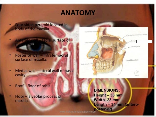
The floor is formed by the alveolar process and if the sinus is of an average size is on a level with the floor of the nose. Without an exam i. Among the diseases are sinusitis frontal sinusitis etmoiditois and. Maxillary sinus floor augmentation also termed sinus lift sinus graft sinus augmentation or sinus procedure is a surgical procedure which aims to increase the amount of bone in the posterior maxilla upper jaw bone in the area of the premolar and molar teeth by lifting the lower schneiderian membrane sinus membrane and placing a bone graft.

Postoperative Stage 2 Panoramic Radiograph Of Left Maxillary First Download Scientific Diagram

Cranio Facial Ct Scan Coronal Sections Left Maxillary Sinus With Download Scientific Diagram

Pns View Showing Destruction Of Floor Of Maxillary Antrum On Left Side Download Scientific Diagram

Intraoperative Endoscopic Views Of The Left Maxillary Sinus Of An Orbital Floor Fracture Through The Antral Window A The Orbital Floor Was Fractured And Periorbital Soft Tissue Was Herniated Into The Maxillary

Emergency Department Computed Tomography Showing A Thickened Inflamed Download Scientific Diagram
Com Sept 2019 Uw School Of Dentistry
Maxillary Sinus Certified Fixed Orthodontic Courses By Indian Dental

Maximum Height Of The Medial Maxillary Sinus Roof Relative To The Download Scientific Diagram

Evaluation Of Haller Cell On Cbct And Its Association With Maxillary Sinus Pathologies Kamdi P Nimma V Ramchandani A Ramaswami E Gogri A Umarji H J Indian Acad Oral Med Radiol

Radiograph Of The Paranasal Sinuses Water S View Reveals A Small Left Download Scientific Diagram

Figure 5 The Prevalence Of Concha Bullosa And Nasal Septal Deviation And Their Relationship To Maxillary Sinusitis By Volumetric Tomography

Figure 6 From Odontogenic Maxillary Sinusitis Need For Multidisciplinary Approach A Review Semantic Scholar

Pdf Case Report Retained Gutta Percha As A Cause For Persistent Maxillary Sinusitis And Pain

