Floor Of The Orbit Panoramic

Dentaltown Where The Dental Community Lives Dental Dental Hygenist Dentaltown

Anatomical Landmarks On Panoramic Radiography Dentstudy Com
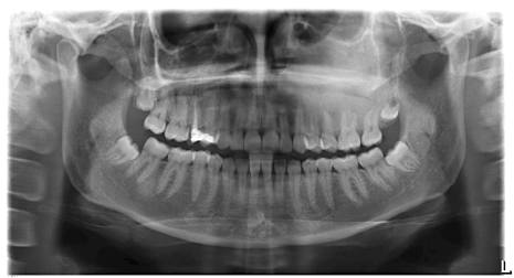
Figure 4

Limited Space Panoramic Outdoor Advertising Floor Graphics

Radiographic Anatomy Of The Skull Organizacao De Estudo

Panoramic Error Recognition Flashcards Quizlet

On a panoramic radiograph the incisive foramen appears as a small ovoid or round area located.
Floor of the orbit panoramic. On a panoramic radiograph the appears as a rounded radiopaque projection of the bone located anterior to the glenoid fossa. External auditory meatus external acoustic meatus. Anatomical landmarks on panoramic radiography enumerate all radiolucent landmarks visible on a panoramic radiograph bony landmarks of the maxilla and surrounding structures. The anatomical landmarks of the are the floor of the orbit and the external auditory meatus.
Preparation to take the panoramic image the clinician needs to have the patient remove jewelry bobby pins hearing aids etc. Only the border of the orbit is visible on most panoramic radiographs. The orbit is a conical structure with its base facing anterolaterally and its apex originating medially as the inlet of all vital neural and vascular structures via the optic foramen superior orbital fissure and inferior orbital fissure. The most frequent example of such a process is the mucous.
The orbit and zygomatic arch. It is also bound by the medial and lateral walls. The upper and lower walls of the cavity are described as the roof and the floor. The anterior rim of the bony orbit the orbital rim is formed by orbital processes from the maxilla z.
Orbit a radiolucent area superior to the maxillary sinus bilateral. Leaving partial dentures in the mouth for a panoramic film will usually obscure important diagnostic information as seen in the above film. Panoramic radiography when large œ and within the panoramic image layer. From the head and neck.
Usually only the inferior border of the orbit is visible over the panoramic radiograph. The forms the floor of the orbit of the eyes the sides and floor of the nasal cavity and the hard palate. The forms the floor of the orbit of the eyes the sides and floor of the nasal cavity and the hard palate. The orbit is a pear shaped cavity with an apex directed posteriorly medially and slightly upward.
Both statements are true. Styloid process a long pointed radiopaque structure that extends from the temporal bone anterior to the mastoid process bilateral. Regarding inflammatory conditions of non odontogenic origin these are usually clearly demonstrated on panoramic radiography if they involve mucosal thickenings arising from the floor of the maxillary sinus. A bony cavity containing the eyeball is a radiolucent compartment with radiopaque borders situated superior to the maxillary sinus.

This Date In Science John Glenn First American To Orbit Earth Earthsky Org John Glenn Earth From Space Nasa Earth

Inner Orbit Lvov Left And Trocto Right By Hybycozo Interactive Art Mirror Modern

Everythingandsome Home House Design Penthouse Living
Https Onlinelibrary Wiley Com Doi Pdf 10 1111 J 1834 7819 2011 01655 X

Pin On Things To Do In London

This Modern New York Penthouse Features Panoramic Views And Sophisticated Decor Stylish Bathroom New York Apartment Sophisticated Decor

Squamous Cell Carcinoma Floor Of The Mouth Panoramic Radiograph Download Scientific Diagram

Panoramic Advertising Format For Warner Bros Pacific Rim At The Metro Centre Panoramic Metrocentre Mallad Outdoor Advertising Metro Center Floor Graphics

30 Modern Corner Windows For Framed And Frameless Panoramic Views Farmhouse Style House Plans Modern Farmhouse Floorplan Farmhouse Architecture

Jp London Md4a134 Solar System Space Removable Panoramic Https Www Amazon Com Dp B008592uh4 Ref Cm Sw R Pi Awdb X T Wall Murals Mural Panoramic

Arounder Travel And Lifestyle 360 Panoramas Castle Panorama Germany

After First Collaborating In 2011 And Again In 2012 Tetra Pak And Orbit Design Studio Reteamed A Third Time To Craft A New Tetra Pak Tetra Office Relocation

Artist S Concept Of Skylab Space Station Cluster In Earth S Orbit Original From Nasa Digitally Enhanced By Rawpixel F Space Station Spacecraft Earth Orbit

